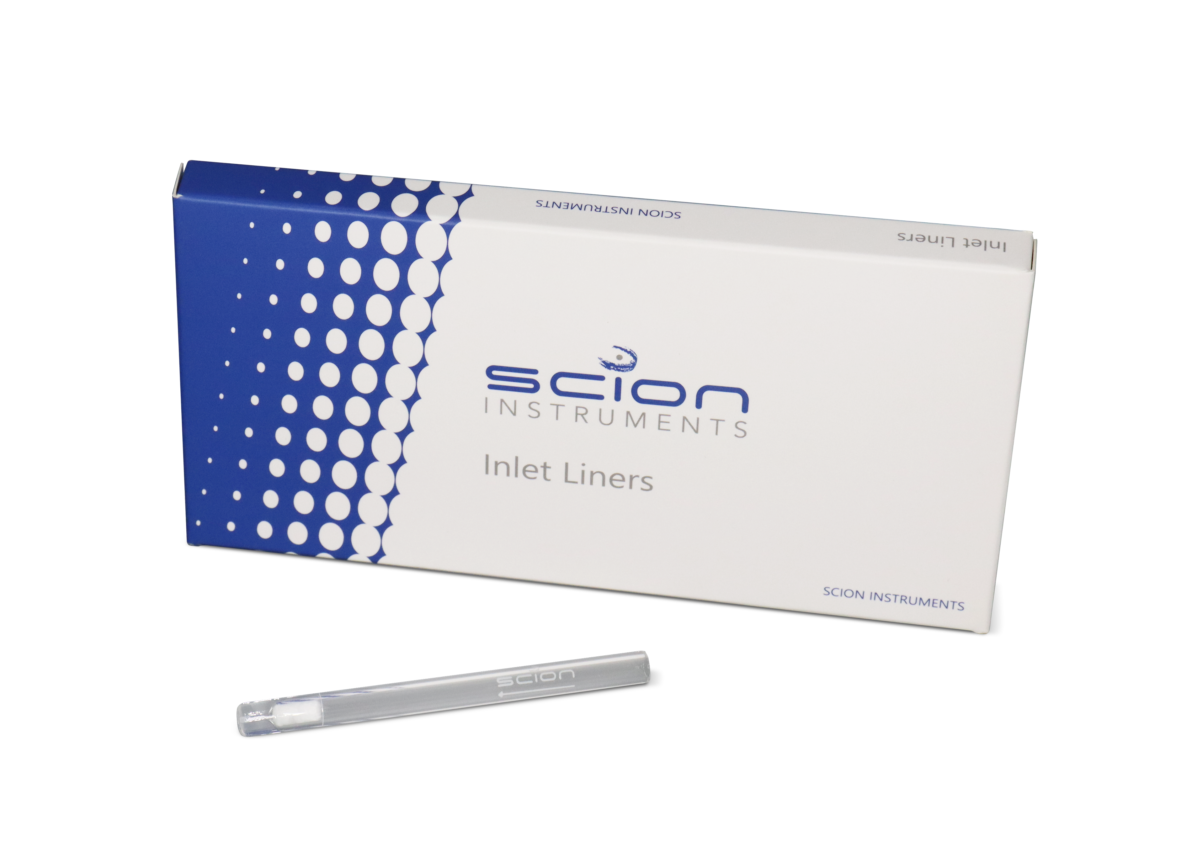
For more detailed information on our GC liners, read our Gas Chromatography Liner Guide.
Gas chromatography liners are integral components in the chromatographic process, playing a pivotal role in optimizing sample injection and vaporization within the chromatograph. These small yet crucial components ensure the accuracy and reliability of your gas chromatography analyses.
GC liners serve as a protective barrier between the sample injection system and the chromatographic column, facilitating the seamless transition of volatile compounds from the sample matrix to the analytical column. They help prevent sample contamination, enhance vaporization efficiency, and contribute to the overall reproducibility of results.
Split/Splitless injector (1177 S/SL)
- SCION qualified inserts use highest quality glass/quartz wool for minimal degradation
- SCION qualified inserts are deactivation treated to minimize activity
- SCION qualified specific geometries for applications
| P/N |
Description |
ID (mm) |
App. |
| 41312100 |
LINER 1177 78.5 L x 6.3 OD x 4MM ID S/SL STRAIGHT QWOOL PK/5 |
4 |
S/SL |
| 41312101 |
LINER 1177 78.5 L x 6.3 OD x 4MM ID S/SL FOCUS PK/5 |
4 |
S/SL |
| 41312102 |
LINER 1177 78.5 L x 6.3 OD x 4MM ID S/SL TAPER FOCUS PK/5 |
4 |
S/SL |
| 41312103 |
LINER 1177 78.5 L x 6.1 OD x 2MM ID S/SL STRAIGHT PK/5 |
2 |
S/SL |
| 41312104 |
LINER 1177 78.5 L x 6.3 OD x 2MM ID S/SL FOCUS PK/5 |
2 |
S/SL |
| 41312105 |
LINER 1177 78.5 L x 6.3 OD x 4MM ID S/SL STRAIGHT PK/5 |
4 |
S/SL |
| 41312106 |
LINER 1177 78.5 L x 6.3 OD x 4MM ID S/SL RECESSED GOOSENECK QUARTZ WOOL PK/5 |
4 |
S/SL |
| 41312107 |
LINER 1177 78.5 L x 6.3 OD x 2MM ID S/SL RECESSED GOOSENECK PK/5 |
2 |
S/SL |
| 41312108 |
LINER 1177 78.5 L x 6.3 OD x 1.2MM ID S/SL STRAIGHT PK/5 |
1.2 |
Split, SPME |
| 41312109 |
LINER 1177 78.5 L x 6.3 OD x 4MM ID S/SL TAPER PK/5 |
4 |
S/SL |
| 41312110 |
LINER 1177 78.5 L x 6.3 OD x 4MM ID S/SL DBL TAPER PK/5 |
4 |
S/SL |
| 41312111 |
LINER 1177 78.5 L x 6.3 OD x 4MM ID S/SL TAPER QUARTZ WOOL PK/5 |
4 |
S/SL |
| 41312112 |
LINER 1177 78.5 L x 6.3 OD x 2.3MM ID S/SL TAPER FASTFOCUS PK/5 |
2.3 |
S/SL |
Split/Splitless injector (1177 S/SL)-Advanced Liners
- SCION Ultra inert liner inserts prevent breakdown of active compounds
- SCION Ultra inert liner inserts realizes best peak symmetry in ‘hot injections’
| P/N |
Description |
ID (mm) |
App. |
| 41312113 |
LINER 1177 78.5 L x 6.3 OD x 0.75MM ID SPME STRAIGHT PK/5 |
0.75 |
SPME |
| 41312114 |
LINER 1177 78.5 L x 6.3 OD x 4MM ID S/SL STRAIGHT QWOOL ULTRA INERT PK/5 |
4 |
S/SL |
| 41312115 |
LINER 1177 78.5 L x 6.3 OD x 4MM ID S/SL TAPER FOCUS ULTRA INERT PK/5 |
4 |
S/SL |
| 41312116 |
LINER 1177 78.5 L x 6.3 OD x 2.3MM ID S/SL TAPER FASTFOCUS ULTRA INERT PK/5 |
23 |
S/SL |
| 41312117 |
LINER 1177 78.5 L x 6.3 OD x 4MM ID S/SL TAPER QUARTZ WOOL ULTRA INERT PK/5 |
4 |
S/SL |
Split / Splitless injector (1177 S/SL) – O-ring inlet Port seals
- SCION Injector O-rings provide best leak tight seal, avoid air leaks
- Graphite or Viton available; Graphite allows higher temperature for inlet (max 450°C)
| P/N |
Description |
| 41312118 |
INJECTOR SEAL O-RING 1177 6.5MM ID VITON PK/10 |
| 41312119 |
INJECTOR SEAL O-RING 1177 6.5MM ID GRAPHITE PK/10 |
PTV Inlet (1078/1079 PTV), On Column Inlet (SPI/1093)
- SCION qualified inserts use highest quality glass/quartz wool for minimal degradation
- SCION qualified inserts are deactivation treated to minimize activity
- SCION qualified specific geometries for applications (i.e. on-column, pseudo on-column or SPME)
| P/N |
Description |
Description (mm) |
App |
| 41312120 |
LINER 1093/SPI 54 L x 4.6 OD x 0.5MM ID COC CONNECTITE R 0.25 PK/5 |
0.5 |
On Column |
| 41312121 |
LINER 1093/SPI 54 L x 4.6 OD x 0.5MM ID COC CONNECTITE R 0.25 PK/5 |
0.5 |
On Column |
| 41312122 |
LINER 1093/SPI 54 L x 4.6 OD x 0.8MM ID COC CONNECTITE R 0.50 PK/5 |
0.8 |
On Column |
| 41312123 |
LINER 1078/1079 54 L x 5 OD x 0.5MM ID S/SL STRAIGHT PK/5 |
0.5 |
S/SL / PTV |
| 41312124 |
LINER 1078/1079 54 L x 5 OD x 3.4MM ID S/SL DOUBLE TAPER FOCUS PK/5 |
3.4 |
S/SL / PTV |
| 41312125 |
LINER 1078/1079 54 L x 5 OD x 3.4MM ID S/SL FOCUS PK/5 |
3.4 |
S/SL / PTV |
| 41312126 |
LINER 1078/1079 54 L x 5 OD x 3.4MM ID S/SL TAPER PK/5 |
3.4 |
S/SL / PTV |
| 41312127 |
LINER 1078/1079 54 L x 5 OD x 2MM ID S/SL TAPER PK/5 |
2 |
S/SL / PTV |
| 41312128 |
LINER 1078/1079 54 L x 5 OD x 0.8MM ID SPME STRAIGHT PK/5 |
0.8 |
SPME |
PTV Inlet (1078/1079 PTV), On Column Inlet (SPI/1093) – Ferrules
- Scion qualified ferrules for PTV Injectors
- Perfect seal on inserts using liner jig, for best precision results
| P/N |
Description |
| 41312146 |
GRAPHITE SEALING FERRULE FOR 1078 & 1079 INJECTORS PK/10 |
Packed Wide Bore – On Column / Flash Inlet (1041/1061)
- SCION qualified inserts use highest quality glass/quartz wool for minimal degradation
- SCION qualified inserts are deactivation treated to minimize activity
- SCION qualified specific geometries for applications (i.e. on-column, pseudo on-column or SPME)
| P/N |
Description |
| 392611943 |
Flash Vaporization Glass Insert for Megabore (pk of 5) |
| 392611944 |
Flash Vaporization Glass Insert for Packed Col. (pk of 5) |
| 392543101 |
Insert, PWOC Injector. Allows installation of 0.53 µm columns |
| 392558891 |
Packed Colum Adapter Kit for On Column, 1/8″ SS Column |
| 392558301 |
Flash Vaporization Column Guide for 3800 |

